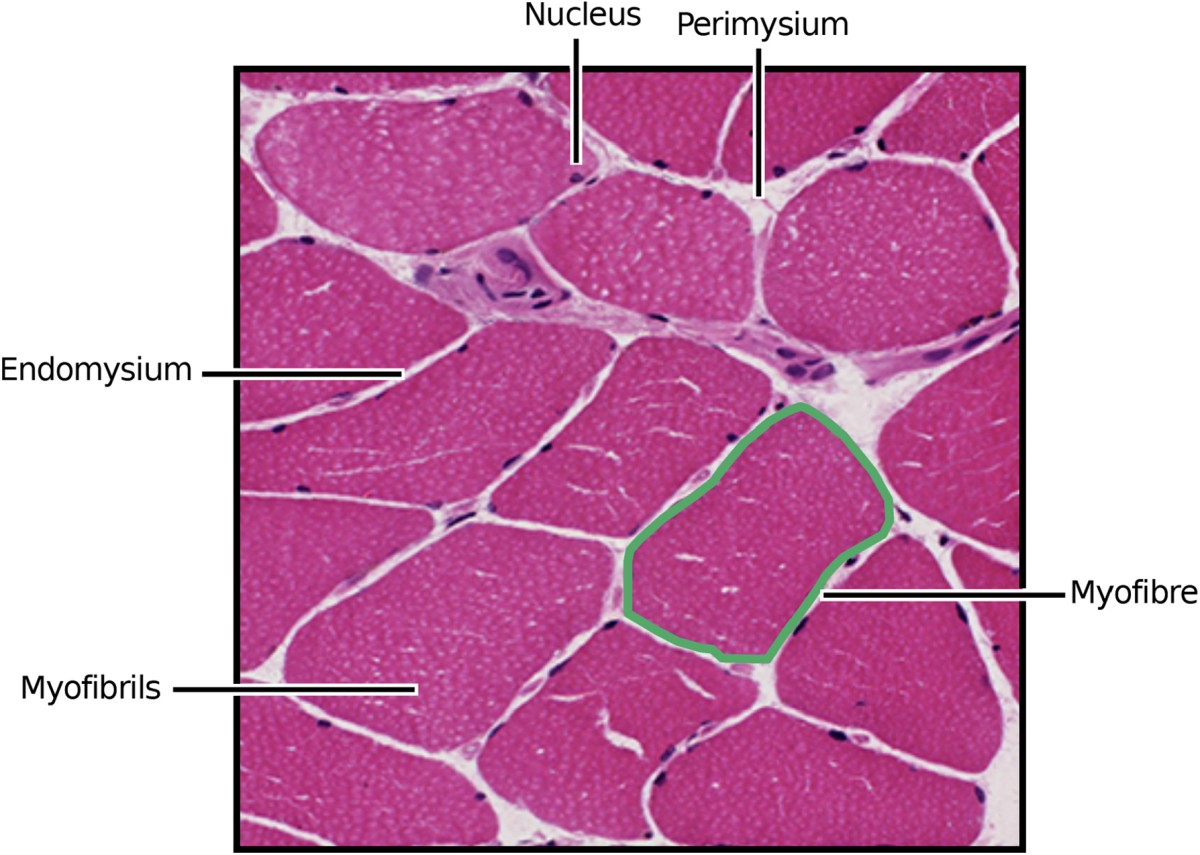
Figure 1

General organisation of skeletal muscle. Polygonal myocytes are shown revealing the presence of peripheral nuclei. The stained sarcoplasm within the myofibrils is light pink. Each fibre is surrounded by mesenchymal matrix known as the endomysium. Myocytes are grouped into fascicles invested by a connective tissue sheath known as perimysium. The perimysium contains nerves and blood vessels that supply the muscle.